Make the internet a better place to learn
Why does the narrowing of the arteries decrease blood flow but increase blood pressure?

Answer:
If you narrow any tube, the flow of fluid will decrease.
Explanation:
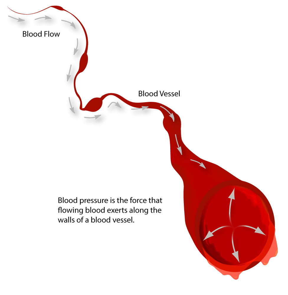
Imagine a rubber tube and think of pinching it. The flow of fluid will slow down. The same occurs in the arteries of the body.
The problem is not as important in the veins as in the arteries.
But since the flow is reduced, how will the body compensate for that? You do need a certain volume of blood flow to the head or you will pass out.
The only way to do this is to somehow increase it by making the heart beat harder and faster.
This will allow the blood to push harder against the tube (vessel) as it does it will expand some. This causes the blood pressure to go up and flow to increase.
This the definition of what we call high BP. "High blood pressure is a common disease in which blood flows through blood vessels (arteries) at higher than normal pressures".
What are premature ventricular contractions (PVCs) of the heart? How are these treated?

Answer:
Premature ventricular contractions (PVCs) are contractions of the heart muscle that originate in the ventricles. They may or may not be treated, depending on the patient's condition.
Explanation:
Normally, the stimulus for the heartbeat is an impulse originating in the sinoatrial (SA) node. But sometimes, a section of ventricular tissue get irritated and start producing their own impulse. When it comes prior to the impulse coming from the SA node, it travels back up the pathway toward the atria.
PVCs can take different forms on an electrocardiogram, depending on where in the heart cycle they occur. But one thing is certain - they are easy to spot, because they look VERY different than what surrounds them.
They can come on their own, in pairs or groups. They may occur regularly or irregularly.
Often, they are benign - no treatment being recommended. But if they do keep occurring, or the patient becomes symptomatic beyond the feeling of the "skipped" beat, then use the ACLS algorithm to treat in the appropriate manner PROVIDED THAT (1) you are properly trained and certified to do so, and (2) it aligns with the protocols under which you operate.
All nervous tissue outside of the central nervous system is part of what nervous system?

Answer:
The NS outside the central nervous system is called the peripheral nervous system.
Explanation:
These are nerves that extend from the brain (cranial nerves) and spinal cord (spinal nerves).
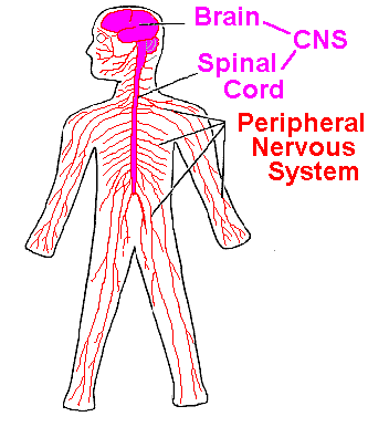
These two systems communicate with each other to make sure our body parts, such as our fingers, can send signals to the central nervous system for processing in our brains.
- Divisions of the Peripheral Nervous System:
There are two functional divisions of the peripheral nervous system the afferent (sensory) division and the efferent (motor) division.
It helps to learn the meaning of some of the words used here. Afferent means to bring into. And efferent means to go out.
Sensory receptors are found at the ends of peripheral neurons and detect changes (i.e. are stimulated) in their surroundings.
Once stimulated, sensory receptors transmit a sensory impulse to the CNS.
 )
)
Motor impulses are carried from CNS to responsive body parts called effectors and a motor impulse is carried on a motor neuron to effectors. There two types:
-
muscles (that contract)
-
glands (that secrete a hormone)
Can the heart itself be damaged by an external force, such as being struck forcefully in the chest? What would be the result of this type of injury?

Answer:
A strong external force can cause bruising to the heart muscle or even cardiac arrest.
Explanation:
Because the heart is a muscle, if an external force acts upon the heart, it can be damaged. One of the most common injuries of this type is myocardial contusion .
This usually occurs because of blunt force to the anterior thoracic wall , such as hitting a steering wheel of a car when not wearing a seatbelt. Often, it is accompanied by rib fractures and punctured lung (pneumothorax).
Patients with a myocardial contusion will present with chest pain, a fast irregular pulse, low blood pressure, and difficulty breathing.
If the contusion is severe enough, it can result in blood building up around the heart ( cardiac tamponade ). This can be life-threatening, because the heart is not able to fill completely and circulate oxygenated blood to the vital organs.
The strongest evidence that cardiac tamponade is present is Beck's Triad : distended neck veins, muffled heart sounds, and low blood pressure. Narrowed pulse pressure and pulsus paradoxus (a drop of blood pressure upon inspiration) are also often present.
One of the other life-threatening complications of sudden force to the heart is a dysrhythmia, an irregular heartbeat.
The force of the impact of a small object such as a baseball, hockey puck, or bat hitting a person's chest with a great deal of velocity can be enough to cause a number of cardiac dysrhythmias, including ventricular fibrillation which would necessitate CPR, defibrillation, and advanced measures.
The most profound injury that can result is cardiac rupture . Blunt force trauma can be strong enough to rupture a heart chamber - this is rarely a survivable injury.
A more detailed explanation can be found in this article from The Journal of Trauma.
What are the layers of the epidermis?
Answer:
The epidermis has either four or five layers (or strata) depending on where it is.
Explanation:
There are two main types of epidermis:
Thin , which is found in places like your eyelids and consists of 4 layers (or strata).
Thick , which is found in areas that experience a lot of wear and tear (like the heels and soles of your feet).
 )
)
The five layers (or four in thin skin) are:
Corneum - This is the outermost, roughest layer layer that consists of 20 - 30 layers of dead keratinocytes. They are dead, flat cells that are filled with a protein called keratin. They flake off the surface of the skin only to be replaced by new cells that rise up from lower layers.
Lucidum - This layer is only present in thick epidermis Lucidum is latin for clear, which makes sense as the Lucidium consists of 2 - 3 layers of clear, flat, dead keratinocytes
Granulosum The first layer to contain living cells, this layer has a grainy appearance due to the cells being moved up as they produce keratin.
Spinosum The cells in this layer look spiny when dried for a microscope sample because of tiny filaments that join the cells together.
Basal/e The bottom layer, this is where mitosis and most of cell production occurs. It also connects the epidermis to the dermis.
A useful, but ironic, way to remember these layers is with the following mnemonic:
Come, Lets Get Sun Burnt
Come - Corneum
Lets - Lucidum
Get - Granulosum
Sun - Spinosum
Burned - Basal/e
I hope this helps, and let me know if I can do anything else:)
What are some functions of muscles?
Answer:
Primarily, muscles are responsible for movement - be this lifting your leg or pumping blood. The movement is achieved through paired contraction and relaxation.
Explanation:
 )
)
There are three main types of muscle which all have different functions. These are:
 )
)
Skeletal - Also known as voluntary or striated (due to stripes caused by muscle fibrils), skeletal muscles are what we generally think of when discussing muscles. Most are joined to bones by tendons and produce conscious movement, such as moving your arm or walking.
 )
)
Smooth - Smooth, or involuntary, muscle are responsible for our bodies automatic processes that we are not conscious of. Smooth muscle is commonly found in the walls of hollow organs such as the stomach, intestines and even blood vessels. It is incredibly important as without it, we wouldn't be able to transport substances (vacoconstriction - movement of blood through vessels - and peristalsis - food moving through the digestional tract - are both caused by smooth muscle), or carry out vital functions, such as breathing.
 )
)
Cardiac This type of muscle forms the walls of the heart and are responsible for it's contraction. It is structurally similar to skeletal muscle, but is controlled by the autonomic nervous system making it involuntary (Thank goodness! - It would be a little difficult to have to remember to make your heart beat!). Cardiac muscle also has the advantage of being very resistant to fatigue.
Hope this helps, let me know if i can do anything else:)
Why does vaccination provide long-lasting protection against a disease, while gamma globulin (IgG) provides only short-term protection?
Answer:
Because vaccination involves active immunity.
Explanation:
Now before we proceed, let us define some terms first.
Let us define the term antigen first. The antigen is like an organism's ID. It needs to be presented before it is recognized. The analogy here is a demon(foreign organism) went to heaven(human body) and in the gate, an ID(antigen) was required. Because the people in the gate saw that the demon had a Hell ID, the Generals(Lymphocytes) were informed what this demon looked like and commanded their subordinates(antibodies) to apprehend the demon.
There are two kinds of immunity:
1. Active Immunity
It is very important to remember that when a foreign immunogenic(capable of inducing immune response) antigen is found in the body, our body, specifically our B lymphocytes, produces substances called antibodies to make it easier for our white blood cells to get rid of the offending organism.
B Lymphocytes, upon antigen recognition, will differentiate into Plasma cells and Memory B cells. Plasma cells secrete the antibody specific to the antigen, and Memory B Cells, as the name implies, will remember that foreign antigen to react quicker in future encounters of the organism with the same antigen, and hence, gives a Long term but slow acting protection.
Example: Vaccination- a part of the organism is injected to the individual therefore stimulating antibody production without causing the disease.
2. Passive Immunity
Passive immunity is administering the antibody itself. And therefore, no memory B cells were produced. However, because the antibodies are readily available, the antigen is neutralized quickly, without the help of B lymphocytes. However, it is only short term because Memory B cells are not produced. Hence, it gives a short term but rapidly acting protection..
Example: Rabies Immunoglobulin is injected to individuals bitten by animals to counter the virus' effects in the body. By the time the body produces antibodies against the rabies virus, the person may already be dead.
Hope this helped! :)
Is the lymphatic system the same as the immune system? Are both of these terms used to describe the same system or are they separate?

Answer:
The organs of the lymphatic system & the immune system are same. But they are functionally separate.
Explanation:
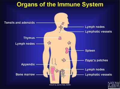
Lymphatic system consists of lymph, lymph vessels, lymph nodes and some organs (tonsils, spleen, thymus, vermiform appendix, Peyer's patches).
The diagram of the lymphatic system :
 )
)
The organs of lymphatic system is the same as the organs of immune system. But these two are different in function.
Lymphatic system has several functions like to drain protein-containing fluid that escapes from the blood capillaries from tissue spaces ; to transport fats from the digestive tract to the blood; to produce lymphocytes and to develop humoral & cellular immunities.
So, the lymphatic system apart from developing immunities, does other things too.
Human body has two major types of Immunities : Innate and Acquired. The humoral and cellular immunities which are developed by lymphatic system is associated with acquired immunity.
The innate immunity is non specific type of immunity and is present by birth. This is not developed by lymphatic system. One example of innate immunity is the presence of HCl (hydrochloric acid) in stomach.
Although these two system share the same organs, functionally they are different.
Here is the organs of the immune system :
Where are the adrenal glands located? What is their function?

Answer:
There are two adrenal glands and they rest on top of each kidney. Adrenal glands secrete hormones : adrenaline, noradrenaline, mineralocorticoid, glucocorticoid & androgens.
Explanation:
Here is a diagram showing the location of adrenal glands on top of each kidney :
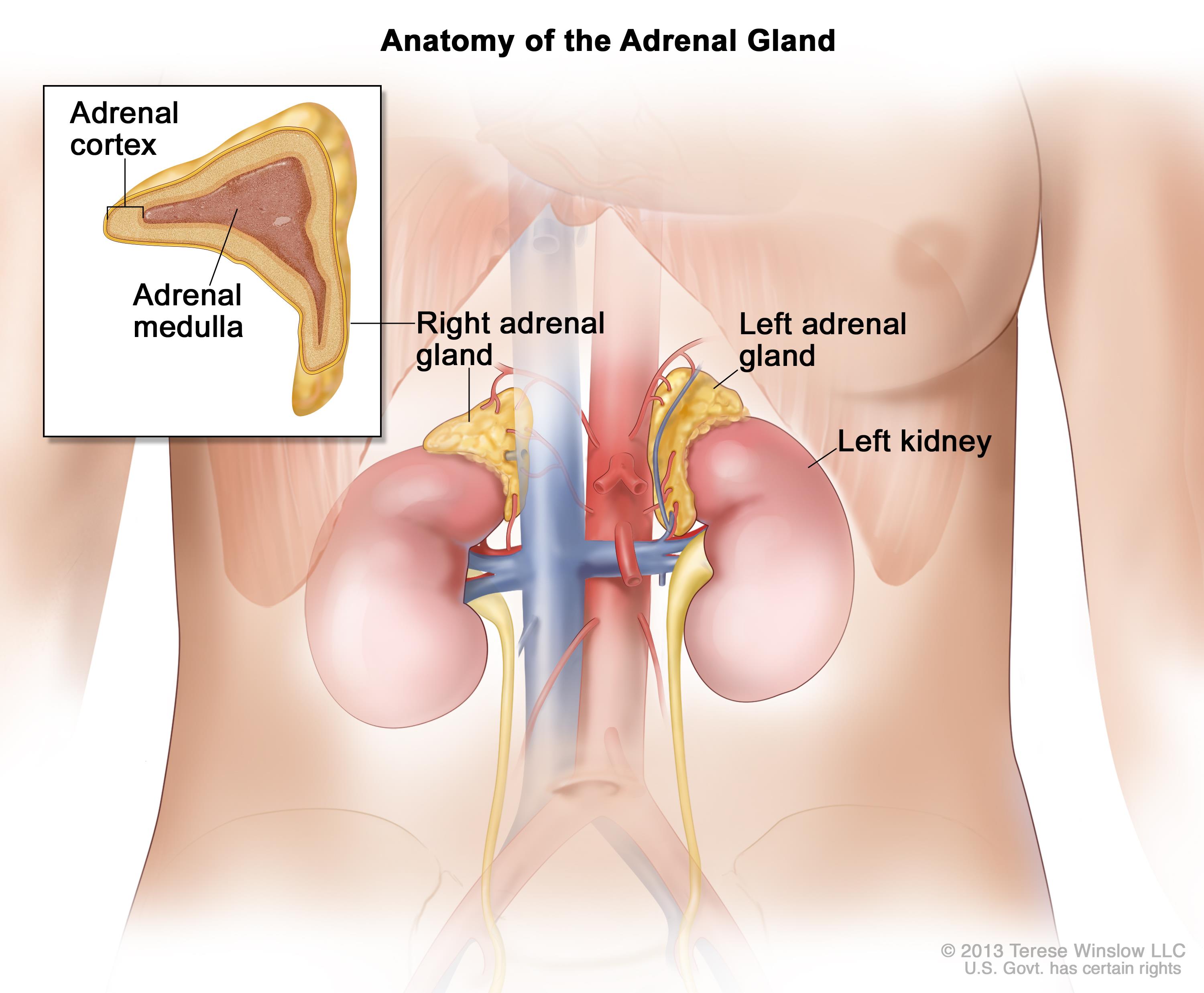
Adrenal gland has two parts, anatomically : adrenal cortex and adrenal medulla. These two act like two separate endocrine glands. The adrenal medulla produces large amounts of the hormone adrenaline, also known as epinephrine, and small amounts of norepinephrine or noradrenaline. Epinephrine and norepinephrine are commonly referred to as the fight-or-flight hormones because they get the body prepared for stressful situations that require vigorous physical activity.
This diagram shows the effects of epinephrine (adrenaline) on different organs of body :
 )
)
The adrenal cortex makes up the bulk of the adrenal gland. Its cells are organized into three layers of cells, forming an inner, a middle, and an outer region of the cortex.
The outer layer of the adrenal cortex secretes a group of hormones called "mineralocorticoid hormones" because they regulate the concentration of mineral electrolytes. The most important of these hormones is aldosterone, which regulates sodium reabsorption and potassium excretion by the kidneys.
The middle layer of the adrenal cortex secretes cortisol, which is a "glucocorticoid hormone". Cortisol stimulates the liver to synthesize glucose from circulating amino acids. It causes adipose tissue to break down fat into fatty acids and causes the breakdown of protein into amino acids. The action of cortisol helps the body during stressful situations and helps maintain the proper glucose concentration in the blood between meals. Cortisol also helps reduce the inflammatory response.
Cells in the inner layer of the adrenal cortex produce small amount of the androgens in both male and female.
Are there two brachial arteries in the body? Are there two brachiocephalic arteries in the body?

Answer:
There are two Brachial arteries but only one Brachiocephalic artery in the body.
Explanation:
This is the diagram of branches of arch of aorta :
 )
)
From the picture we can see arch of aorta has three branches : brachiocephalic artery, left common carotid artery and left subclavian artery.
The brachiocephalic artery is in right side and shortly after its commencement it gives rise to right common carotid artery and right subclavian artery.
Whereas in the left side, the left common carotid artery and the left subclavian artery arises directly from the arch of aorta. So, there is no left brachiocephalic artery. And we are left with only one brachiocephalic artery.
The right and left subclavian arteries then supply right and left arm by forming right and left axillary arteries. The axillary artery in each arm then gives rise to brachial artery. Yes there are two brachial arteries. One in each arm.
Here is a couple of pictures. First one shows how brachial artery is formed. The second one shows two brachial arteries :
 )
)
 )
)